Think of it like a kidney stone, but in your mouth. When material builds up over time, the salt in the saliva glands calcify and form a stone in the glands of the mouth called a salivary stone. Or, if you want to be fancy, the medical term is sialolith. When it blocks the salivary glands, it is called sialolithiasis.
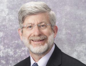
Salivary endoscopy for salivary stones was the November 3rd webinar topic hosted by The Eye & Ear Foundation with presenter Dr. Barry Schaitkin, Professor, Department of Otolaryngology, Emeritus Program Director, Otolaryngology Residency Program at the University of Pittsburgh Dept of Otolaryngology. 
Back in 1988, at a national conference, Dr. Schaitkin attended a presentation on salivary endoscopy, which he had not heard of. He thought it was the most interesting minimally invasive technique for what was at the time a disfiguring operation. When he returned, he told the department chairman that they had to start doing it. The FDA didn’t approve it until 2005. At that time, Dr. Schaitkin went to Europe for instruction and training and was one of the first people in the U.S. to start using it when it was approved.
Symptoms and Presentation
Salivary stones are rare, affecting only one out of 10,000 people. Not much is known about its cause, but it is not related to water hardness or calcium intake. Most will show on x-rays.
Classical presentation symptoms include recurrent swelling, especially during meals. Acute episodes are associated with inflammatory signs. Diffuse swelling can occur on one or both sides. An inability to express free flow of saliva is one sign, and stones may be palpable on examination.
Most of the time, Dr. Schaitkin said, saliva over time can be slowly expressed into the mouth so patients are better and only miserable when they are eating. But the whole system can get infected due to the lack of flow.
One big difference between kidney stones and salivary stones is that the latter is more visible.
Imaging and Removal
A sialogram can be used if there is no acute infection, but it is challenging to perform and read. It involves putting an IV catheter in the mouth and injecting dye. MRI sialography is used with more complicated cases. A virtual salivary endoscopy uses an endoscope.
The opening where saliva comes out is extremely tiny; it only must be big enough for water to run through it. So, the challenge is designing an instrument that can fit through the channel and extract the stone. Micro instruments tend to break if they grab the stone.
There are different instruments of varying sizes that are used for dilation and extraction. The stone – usually yellow – cannot be collapsed with fingers or an instrument edge.
Sialendoscopy is challenging. The rigid scope is delicate and has a steep learning curve. There is risk of perforation, vascular injury, and stenosis. Unless the physician is experienced at doing a lot of this procedure, it can be difficult to get the scope in, find the stone, and then deliver it. Dr. Schaitkin said he is lucky since the department sends him cases, along with community doctors in eastern Ohio, Western PA, and even further away.
Procedures
There is no correlation between the degree of gland alteration and number of infectious episodes or duration of symptoms. Less than 50 percent of removed glands for swelling are histologically normal. In the past, patients had scar tissue and lost function of their salivary glands. The goal of salivary endoscopy is to remove the obstruction and preserve the salivary glands.
If the stone is small, it can be removed with local sedation. If the stone is large, extracorporeal shock waves – which have been around since 1986 – can be used to break it into smaller pieces. The problem is the device is so powerful that when put near the face, most fillings fall out. It is also so loud that it can result in hearing loss.
Two companies in Europe invented a very small version of this that is used in the office without anesthesia and can sit on the skin. Unfortunately, it is not available in the U.S. Dr. Schaitkin said it is not likely it will ever be FDA-approved, because there is not a market for enough of these devices to be sold in the U.S. to make it profitable to go through the FDA process. Canada does not have it either. Patients go to Europe for this technique.
What the U.S. does have are laser procedures. The same laser used for kidney stones, in fact. Pieces are extracted one at a time. But there is a love-hate relationship with the laser. The scope does not like lasers very much.
“When the laser strikes the stone, it produces a clone of a chemical where the heat interacts with the stone,” Dr. Schaitkin explained. “If it’s too close, the chemical will adhere to the tip of the scope, so it impedes vision and has narrowed the working channel where the instrument goes, to the point where these very very delicate, precise instruments that just fit through these channels no longer fit. Vision goes away and instruments can’t work as well.”
These telescopes, by the way, cost about $7,000 apiece, so it is not good when one becomes non-functional during a case. A lot of technique must be employed to prevent injury.
Size Matters
If the salivary stone is nice and small and floating in the channel, less than 3-4 millimeters in size, it can generally be removed very minimally. Medium sized stones – depending on which gland they are on – may be amenable to laser technique or a combination of a mouth incision and endoscopic technology.
Very large stones require a hybrid approach in removing the stone completely. This means a large incision in the mouth. If it is in the parotid gland by the ear, hooks and retractors are used to pull facial soft tissue forward, which is a more involved process. The nerve that makes facial muscles move runs through the middle of this gland. Doing the procedure this way ensures that the gland stays in place, with its function preserved and no chance of complete facial paralysis.
After stone removal, all patients have an endoscopy to make sure there are no stone fragments or other stones present. The system is irrigated and checked to make sure the closure is tight.
Q&A
Unlike kidney stones, salivary stones do not have any dietary adjustments that people can make. There are also no health predispositions to salivary stones. After it is removed, patients have about a five percent chance of getting another stone in their lifetime – but this is not very common. Dr. Schaitkin encourages people to stay well-hydrated. If you have some relatively sluggish glands, massage the gland after meals. There is nothing you can do to prevent salivary stones.
Recovery time increases with the size of the stone. A basic salivary endoscopy means just recovering from the anesthesia. There is no post-operative medicine or pain. A very large stone and incision means being uncomfortable for three to five days. Patients are not back to normal for a week or even two, but only off work for about three days. For patients whose stones have to be approached from outside the face, it takes five to seven days until they are back to full function.
There is no correlation between head and neck cancer with or without radiation and development of salivary stones. But head and neck cancers are unkind to salivary glands. “Salivary gland function is probably the first thing that head and neck cancer patients miss,” Dr. Schaitkin said. A permanent loss of function occurs in some cases. A fair bit of work is being done around the country with STEM cells and trying to recover salivary function after radiation treatments. Dr. Schaitkin thinks there will be something available within the next five to 10 years.
Salivary stones are seen in all races and ages, including children. Numbers are slightly higher in Europe than in America and Asia, but not dramatically. Peak cases are seen in 30-60-year-olds.
Visit https://eyeandear.org/donate to support our research and educational efforts. Please register for the mailing list to stay informed on our research and patient care advances. Should you have any questions please email Craig Smith at craig@eyeandear.org.