S+S, Fall 2023
Only six devices in the world house a rare kind of imaging technology called laser doppler holography. Three are in Paris and two are in other parts of France. Ethan Rossi, PhD’s lab, Rossi Lab, or the Advanced Ophthalmic Imaging Lab, has the only one in the U.S. and the only one outside of France. The Eye & Ear Foundation helped fund this through a generous gift from Dr. E. Ronald Salvitti, an alumnus of the University of Pittsburgh School of Medicine and an Eye & Ear Foundation Board member.
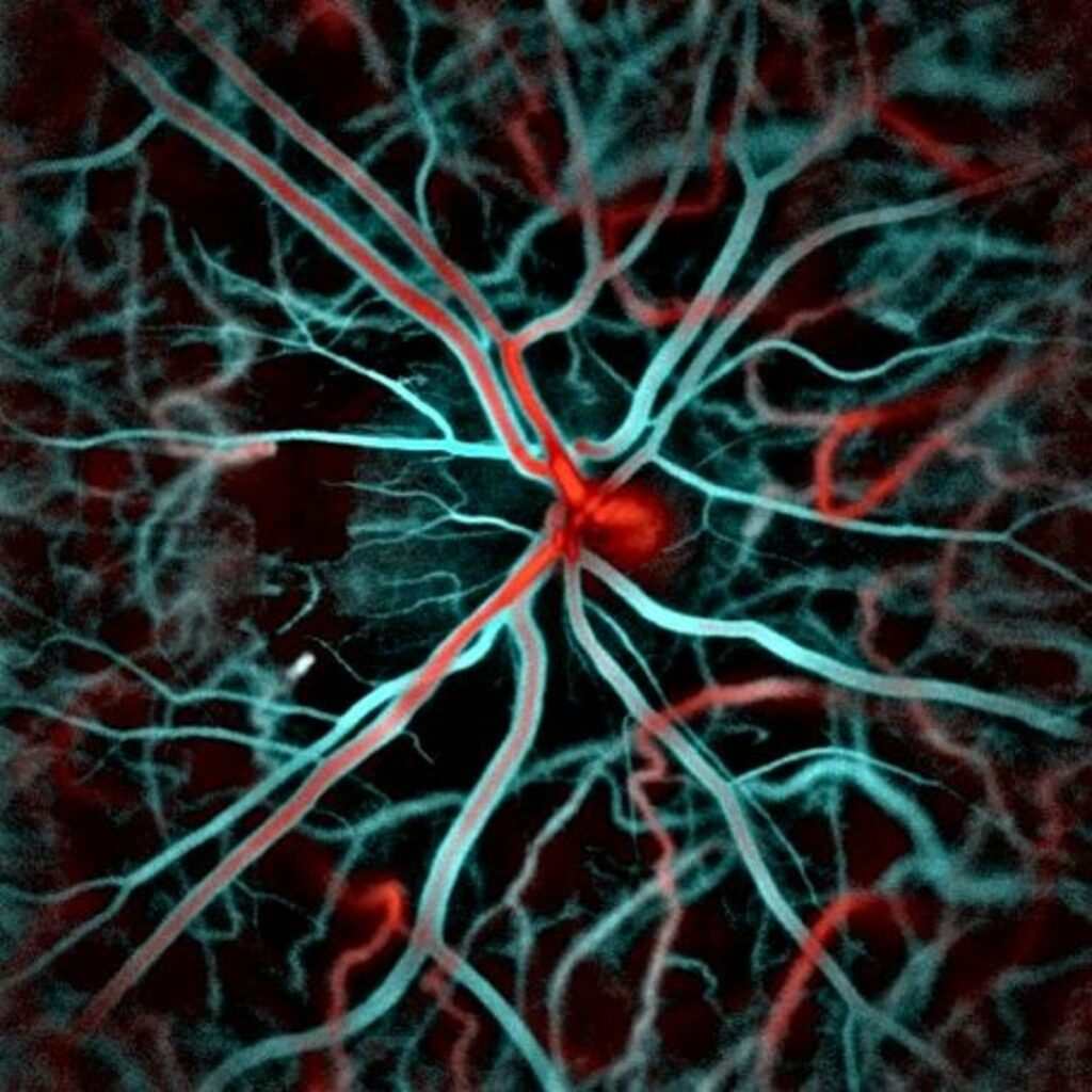
“This is a type of imaging technology that allows us to see functional activity in the retina with high precision,” Dr. Rossi said. It relies on the principle of interference for generating the images. The technology is similar to optical coherence tomography (OCT), which is commonly used in the clinic. The difference is the way the images are acquired. With this new technology, images are received at a very fast rate, which means the ability to view rapid changes over time. “We think the technology has a lot of potential applications beyond what we’ve been starting to look at,” Dr. Rossi said.
The focus has been looking at vascular activity in the retina. The technology generates images that show where the blood vessels are as well as about the blood flow. A novel feature of the technology is that arteries can be identified from veins just from the direction of the doppler signal. Different types of vascular changes are accessible, so the imaging can be used to understand how intraocular pressure/glaucoma affect the blood flow. Not all of these things could previously be done, even with the use of contrast agents injected intravenously.
Dr. Rossi explained how the device works: light from a laser is split into two paths. One path sends light into the eye, while the rest of the light goes directly into the camera, serving as a “reference beam.” The light that comes out of the eye interferes with the reference beam. It is that interference that provides a signal about the activity of what is happening inside the eye. These interference patterns, called interferograms, are captured at a very fast rate. The camera runs at 67,000 frames per second and is one of the fastest cameras in the world. A “slower” camera, which runs at 10,000 frames per second provides a real-time image and streams that data to the computer for the experimenter to evaluate the data during acquisition.
The light-sensing part of the eye, the retina, is one of the most metabolically active tissues in the body, which requires a lot of blood flow. “Evaluating blood flow in the eye can tell us a lot about what’s happening in different diseases, many of which have a vascular component,” Dr. Rossi said.
Dr. Rossi wants to look at changes in patients that have age-related changes to the retina, like age-related macular degeneration. “I’m interested in the potential to use this technology to be able to detect earlier changes than we typically can,” he said.
Dr. Rossi is trying to secure funds to have dedicated personnel on this equipment and support to bring this technology forward.