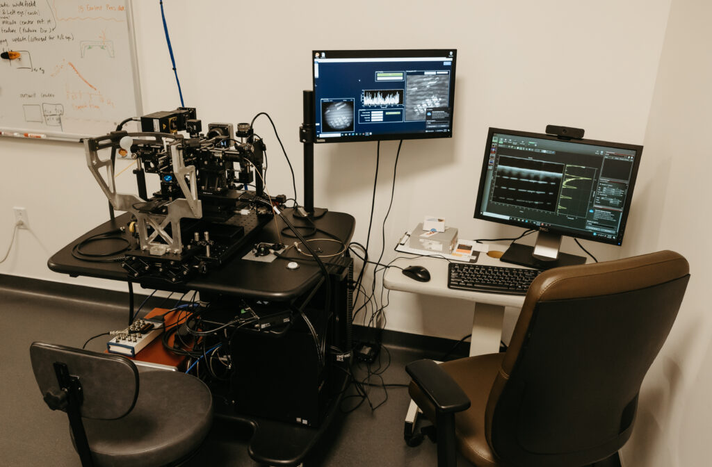
Why are we putting so much effort into figuring out how to image the retina down to the microscopic scale, asked Ethan Rossi, PhD, Associate Professor of Ophthalmology, Assistant Professor in the Department of Bioengineering, University of Pittsburgh Swanson School of Engineering, in the Eye & Ear Foundation’s first webinar of the year. On January 13, Dr. Rossi was one of the presenters for “The Role of Advanced Imaging Technologies in the Future of Eye Care.”
The answer to Dr. Rossi’s question is that current clinical imaging lacks the resolution to see single cells. Clinical instruments show us the structure of the retina at the tissue level. New treatments aim to restore vision through gene or cell-based therapies. The imperfect optics of the human eye limit image resolutions in conventional imaging systems. Therefore, new tools are needed to image the cells in the eye and track their changes in response to treatment.
Principle of Adaptive Optics Ophthalmoscopy
Adaptive optics technology corrects imperfections, or aberrations, in the eye’s optics. Aberrations are measured with a wavefront sensor and corrected with a special mirror that can change its shape to undo the distortions caused by the optics of the eye. When the adaptive optics are turned on, the optics are corrected, making the cells visible, and resolution is improved.
With multi-modal adaptive optics ophthalmoscopy, various properties of light are used to study different cells in the eye. Light reflected out of the eye shows the light sensing cells, fluorescence is used to image another class of cells, while the phase changes that the light undergoes provides an additional way to show cell contrast, revealing immune cells and blood vessels.
Inflammation in Inherited Retinal Degenerations (IRDs)
- IRDs are caused by mutations in genes primarily expressed in photoreceptors and RPE cells
- Many gene therapies for IRDs are currently in clinical trials
- This revolution in therapeutic development has caused a reemergence of interest in the importance of inflammation in IRDs as there is now widespread recognition of gene therapy associated inflammation
- Can advanced imaging provide deeper insight into the inflammatory process in IRDs?
- What biomarkers are accessible to our advanced imaging tools in IRDs?
- How can we use imaging to better select patients for clinical trials or to monitor patients undergoing gene therapies?
These types of tools are broadly applicable to other conditions as well, as many different diseases have an inflammatory component, like glaucoma and age-related macular degeneration.
Microglia and Inflammation
- Microglia are the resident immune cells of the retina
- Form a network of cells that can be activated during inflammation
- Can be triggered by inflammatory responses to pathogens and tissue trauma and pave the way for the recruitment of other immune cells into the eye
- Microglia play an important role in numerous retinal diseases, including uveitis, glaucoma, optic neuritis, AMD, and IRDs
Adaptive Optics Scanning Light Ophthalmoscopy (AOSLO)
Multi-modal imaging is essential to see all the different changes that occur during inflammation in retinal degenerations. The different cells in the eye also vary in size across the retina, with the light sensing cells being smallest in the area called the fovea at the center of our vision.
This area is challenging to image, Dr. Rossi said. In some patients, there are few structural details visible in this area, aside from cystic changes that are very clear to see in one of the imaging modalities, which is why this technology is so beneficial.
Ideally, the patient is seen before treatment, and then monitored after with this imaging to evaluate the efficacy and monitor the changes to the cells over time..
Other types of image processing can be done to highlight the cysts better. “We are working on this so we can segment and quantify these,” Dr. Rossi said.
Summary & Conclusions
- Inflammatory biomarkers include microcysts and immune cells in areas of active degeneration
- Microcysts challenge cone detection, but cones can be seen with appropriate focusing
- Loss/remodeling of fluorescence signal suggests loss of RPE cells
- Autofluorescent cones may be a biomarker of cones undergoing stress on the path to degeneration
- Multi-modal imaging is essential for understanding the complex mix of cellular structures accessible to AOO in patients with IRDs
- Advanced imaging can allow for careful monitoring of inflammation over time at a cellular level in the living eye
In terms of ongoing and future work, Dr. Rossi said his lab is working on being able to see additional structures and existing ones better, in part by improving the image processing. One of these goals is to better detect changes over time, with shorter intervals between images to capture faster moving cells.
Fully Automated OCT Imaging for Ex Vivo and In Vitro Tissue Screening
Researchers at the Vision Institute, led by Dr. Shaohua Pi, are pioneering a groundbreaking fully automated Optical Coherence Tomography (OCT) system designed to transform tissue screening for drug discovery and disease modeling. This innovative platform is optimized for high-throughput, non-invasive imaging and analysis of 3D tissue structures, offering unprecedented capabilities for real-time monitoring and quantification.
The Challenge
Traditional tissue analysis relies heavily on destructive histology methods, which lack the ability to provide continuous, real-time insights into tissue dynamics. This limitation hampers high-throughput applications critical for drug discovery and tissue engineering. To address these gaps, Dr. Pi’s team developed an advanced OCT system tailored for ex vivo and in vitro applications, including retinal organoids and explants.
System Highlights
The fully automated OCT system features a motorized sample manipulation stage with a high field of view, coupled with computer vision algorithms, including Single Shot Multibox Detector (SSD), for precise object detection, and advanced tissue and inner structure segmentation model, for comprehensive and reliable readouts. This setup ensures rapid and accurate sample handling, with detection success rates of 100% and response times as low as seven milliseconds.
Key features of the system include:
3D Imaging and Quantification: Captures high-resolution, volumetric data, enabling detailed assessments of tissue architecture and cellular organization.
Deep Learning Integration: Employs advanced segmentation algorithms like Multi-Kernel Receptive Field (MKRF) and Adaptive Dual Attention (ADA) to differentiate complex tissue types, achieving exceptional segmentation accuracy (e.g., AROC=0.99 for certain tissue classifications).
Validated Metrics: Provides robust readouts such as tissue volume and thickness, with validation metrics like Z’-factors (>0.5) and SSMD (β > 2), which highlight the system’s reliability for drug screening applications.
Advancing Drug Discovery
The team’s system has demonstrated its potential through preliminary studies on retinal organoids and explants. Using models of retinal degeneration, the platform identified significant photoreceptor preservation and tissue thickness improvements with positive treatment responses. This capacity to non-invasively monitor therapeutic efficacy in real time represents a major advancement in preclinical testing.
Dr. Pi emphasizes the significance of the platform: “Our OCT system bridges the gap between traditional tissue analysis and the demands of modern drug discovery. By providing scalable, high-throughput imaging and analysis, we’re opening new doors for therapeutic innovation, particularly in retinal and neurodegenerative diseases.”
Collaborative Efforts
The project benefits from the expertise of collaborators Dr. Yuanyuan Chen and Dr. Susana da Silva within the department, who are contributing to the validation and application of the platform for tissue-based drug discovery.
Outlook
As the team continues to refine the system, the fully automated OCT platform is poised to become an indispensable tool for researchers worldwide, enabling breakthroughs in personalized medicine, regenerative therapies, and beyond. With its ability to deliver scalable and dynamic tissue analysis, the system stands to redefine standards in biomedical research and clinical diagnostics.
For further information, contact Dr. Shaohua Pi at shaohua@pitt.edu or visit the UPMC Vision Institute website.
Object detection (computer vision) with a single shot multibox detector (SSD) has 300 images for training and test in 7 milliseconds with 100% success rate of detection.
Take Home Message
- We are working on a fully automated OCT system for high throughput tissue screening
- Our system enables scalable, non-invasive, and real-time tissue readouts and analysis
- The readouts are based 3D characterization and can capture the dynamic behaviors
- We have preliminary validated the system in retinal organoids and explants for potentiation therapy discovery
