Dr. Ken Nischal, MD, FAAP, FRCOphth, opened the Eye & Ear Foundation’s February 28, 2025, webinar, “Vision and Down Syndrome,” by pointing out how apt it is that World Down Syndrome Day was exactly three weeks away on March 21. The date was chosen because March is the third month and Trisomy 21 means an extra chromosome.
Dr. Nischal has impressive credentials: a pioneer of pediatric corneal transplants, he is also Executive Vice Chair, Department of Ophthalmology; Division Chief, Pediatric Ophthalmology and Strabismus; Medical Director, Digital Health UPMC Children’s Hospital of Pittsburgh; Assistant Medical Director, UPMC International Medicine; Professor of Ophthalmology; Co-Executive Officer and Co-Founder, World Society of Pediatric Ophthalmology and Strabismus (WSPOS).
He started off with some history. Down syndrome was first described in 1846 by Edouard Onesimus Seguin in France. The next description was by John Langdon Down in Surrey, England, 1866. The chromosomal basis was discovered in 1959 by Jerome Leleune (or Martha Gautier?). Martha worked in Leleune’s lab and likely did not get the credit she deserved, so Dr. Nischal wanted to put her name out.
Ocular problems in Down syndrome (DS) fall into two specific categories: visual function and anatomical ocular problems.
Visual Function
Eighty percent of children with DS have significant refractive error, where they are not seeing properly. This becomes more obvious as they get older due to failure of emmetropization, a vision dependent ocular phenomenon that directs the growth of the eye. The eye tries to grow to a point that is genetically influenced but can also be environmentally influenced. If emmetropization does not exist or is not working properly, their eyes become more hyperopic (more plus) or more myopic (more minus). “That’s what we think happens in DS patients,” Dr. Nischal said.
A couple of decades ago, a woman named Margaret Woodhouse raised the issue that children with DS seem to underfocus for near work or targets. This means developing a blurred image when looking at things close up. If this is not corrected by the time you are eight, that blurred vision is uncorrectable.
Hypo-accommodation can present in various ways in children with DS. About 55-70% have it. Strabismus – when the eyes are not aligned – is also common. If it is not corrected, the brain gives up on the image coming from the eye. Children with DS also have a higher incidence of nystagmus and brain-based visual impairment (CVI).
Prevalence and Type of Strabismus
Pseudoestropia occurs when there is an extra fold of skin, a characteristic feature in Trisomy 21. Strabismus occurs in about 25-40% but can be as high as 60%. Nystagmus has about a 10-30% prevalence. Accommodation insufficiency is common, along with near vision issues.
Sometimes when a child is given glasses, their eyes straighten. If they do not, it can be surgically corrected. The big issue is that if a child with DS has a severe developmental delay, any amount of surgery done is controversial. If you get it perfectly right, there is evidence that the eyes will begin to drift out, so the surgical dose is questionable. Some people are beginning to use botulinum and toxin, a paralyzing agent, injected in the muscle pulling the eye.
Nystagmus
Nystagmus is a rapid, repetitive eye movement that can be horizontal, vertical, or torsional, or a combination. It occurs in 30% of patients. Sometimes children will have a null position, where they hold their head in a particular position because in that position, they have the least blur. The cause of nystagmus is unclear, but recent papers have suggested that the fovea – a very central part of vision that fixates it – is not normal. An optic nerve anomaly may also be a cause.
One study found that about a quarter of all children with DS had Abnormal Head Posture (AHP). Having strabismus of any kind, and particularly incomitant strabismus, nystagmus, or both, is highly correlated with the development of an AHP. Almost 19% of DS patients with AHP had no definitive cause that could be determined.
An important thing to know, Dr. Nischal said, is that sometimes you can see AHP in patients with DS because they are notoriously known to have alanto-occipital instability. This is when ligaments that attach to the base of the skull are a little lax. Sometimes if a patient with DS suddenly develops a head posture and it looks like their neck is in spasm, it could be an atlanto-occipital dislocation. This is not common, but before most patients with DS have any kind of general surgery, an exam is done to make sure the atlanto-occipital is stable. This is usually done clinically or with an x-ray to make sure no damage will be done while they are asleep.
BBVI (CVI) in DS
Margaret Woodhouse spent a large part of her life looking after children with DS. She was an optometrist who wrote a paper with her group about how they looked at a group of children and used an Insight Inventory by Dr. Gordon Dutton. It has 52 questions referring to visual perception and visual motor difficulties across six domains: visual search, visual fields, visual attention, perception and movement, visual guidance of movement, and recognition and navigation. The caregiver rates whether or how the question is present in their life using a four-point scale. Tailored habilitation strategies are then created and shared with the family, caregiver, and school.
Five of the questions were taken as a “screening tool” for CVI. Using this screening tool, 38% of the children in the survey were positive for suspected CVI.
Parents are asked if their child has difficulty walking downstairs, finds uneven ground difficult to walk over, and has difficulty seeing things which are moving quickly (like small animals). People with BBI have difficulty with these, and it has nothing to do with their ability to use the two eyes together.
At Dr. Nischal’s clinic at Children’s Hospital of Pittsburgh, the color of the corridor is different than the color of the floor in each room. If he sees a patient with DS walking in and they suddenly stop at the threshold, this tells him there may be a processing problem. He then reduces the number of doctors, students, residents, and fellows coming into the room, because that affects how compliant the patient will be. If they are disoriented and confused, what is called crowding of a visual scene, they can get anxious. “This is something worth knowing,” Dr. Nischal said. “If you are taking someone out for a walk who has DS, try to make sure that where you’re walking is sort of even, not across too many steps, and hold their hands so they feel comfortable.”
Anatomical Characteristics of DS
- Upward slanting of eyelid fissure (60-80%)
- Large epicanthal folds (31-97%) – can be a terrible problem, lid margins can get very crusty
- Blepharitis/inflammation of the eyelid
- Epiblepharon
- Nasolacrimal duct obstruction
- Brushfield spots on the iris
- Cataracts (4-30%)
- Keratoconus (a cone-shaped distortion of the cornea/front layer of the eye) (<15%)
- Optic nerve anomaly (rare)
- Glaucoma (increased eye pressure) (rare)
The American Academy of Pediatrics has recommendations for eye exams in patients with DS:
- Within the first 6 months of life
- Every year from 1-5 years
- Every 2 years from 5-13 years
- Every 3 years from 13-21 years
- Follow up can be more often if needed
What they do not talk about are the adults. Dr. Nischal thinks adults with DS should be seen every year because they can run into problems that can affect their whole function.
Keratoconus and Developmental Delay
Dr. Vishal Jhanji, MD, FRCS, FRCOphth, FARVO, corneal specialist and clinician scientist, Immediate Past President of the Eye & Contact Lens Association; Vice Chair, Quality; Director, Cornea Service; Professor of Ophthalmology; Co-Director, Clinical Trials; and Clinical Co-Director, Funderburgh Cornea Regeneration Project, talked about keratoconus. He is proud that today Children’s Hospital of Pittsburgh is one of the best centers for providing keratoconus care. He takes care of adult patients with DS, which is slightly different than what is done at Children’s for the pediatric population.
Keratoconus is characterized by axial/paracentral stromal thinning, progressive irregular astigmatism, and usually presents in the second decade of life. At a very young age, patients or kids will start complaining about the change in their spectacle prescription. This might not happen in every patient who has a developmental delay. “If we are not careful, especially in a population such as DS, we might miss it for a long time until it is too late to take good care of these patients,” Dr. Jhanji said.
In a normal cornea, the cornea stroma is made up of parallel collagen. There are bridges, or “cross-links” between collagen. In keratoconus, there is a reduction in the number, degradation, displacement, and rearrangement of collagen fibers. This presents clinically with irregular myopic astigmatism, epithelial iron deposits, corneal scarring, or acute corneal hydrops. Early signs are Fleischer’s ring and Vogt’s striae. Late signs are Munson’s sign, Rizutti’s sign, or a corneal scar.
Machines map out the corneal surface and can show the different stages of disease. Advanced software can show whether a patient has keratoconus. The actual biomechanical properties in keratoconus can be measured. It is very difficult to ask a patient with DS to sit on a machine and focus on a tiny beam of light so they can be scanned and measured.
Twenty years ago, treatment for all keratoconus patients used to be glasses, and if that did not work, they were offered corneal transplantation. This is challenging for people taking care of patients with a developmental delay, because while it may not be that difficult to perform a corneal transplant in a patient with DS, the real problem is post-operative care and follow up. This is one thing that made Drs. Jhanji and Nischal work harder on treatment protocol, such as how surgery is done, what education is provided to parents and other family members, and how frequently patients are to be seen.
Fortunately, the FDA approved a treatment called collagen crosslinking.
Collagen Crosslinking
Riboflavin mediated crosslinking frameshifts a biomechanically weak cornea to a safer biomechanical level. Basically, the cornea becomes a little brittle and keratoconus stops progressing. The skin of the cornea is removed, and vitamin eye drops (riboflavin) are used for half an hour. Then a UV light is shone through a lamp onto the patient’s eye for another half hour. It is straightforward, and results are very encouraging. Most previous studies have shown that it is very effective in more than 80% of patients.
Optical coherence tomography is another kind of imaging modality, in this case to tell whether the riboflavin (yellow in color) has actually penetrated to a point where it is supposed to before the cornea is treated with UV light.
Another benefit: collagen crosslinking is cost effective compared with standard management at an incremental cost of 3174 Euro per QALY in the base case, which reduces the need for corneal transplantation. This is especially relevant for patients with DS, as this saves a lot of money for patients who do not need surgery or follow ups for the rest of their lives.
Studies have shown that failure to deliver crosslinking to developmentally delayed individuals (especially Trisomy 21) results in marked progression of keratoconus.
Barriers for crosslinking in children or developmentally delayed adults include:
- Holding still for an hour and 10-20 minutes
- Potential to rub the eye overnight
- Increased risk of infection (because the corneal skin has been removed during the surgery)
- Regression worries – eye rubbers
These patients have to be followed up from time to time because they can progress later in life. Even though the success rate is 80-85%, the 10-15% of patients who might progress or need another crosslinking must be taken care of.
Experience
At UPMC Children’s Hospital of Pittsburgh, Dr. Nischal is the surgeon for patients with DS. He has done 30 eyes of 21 patients with developmental delays. The mean age at presentation was 23 years, though disease onset may have been much earlier.
Solutions to the challenges presented by patients include general anesthesia, intraoperative OCT to assess the soak of riboflavin, aggressive pre and post management of inflammation (namely allergic conjunctivitis), and behavioral therapy for eye rubbing.
A clinical exam is attempted to look for clinical signs, though it may not be possible to get a scan to measure corneal thickness while in the clinic. Ultimately, all these patients need an exam under anesthesia.
After crosslinking surgery, it is important that patients do not rub their eyes. Surgeons create a large scratch to make sure the crosslinking technique is effective. They temporarily close the patient’s eyelids (tarsorrhaphy) with sutures while they are asleep; these are very easy to remove in the clinic. This provides ample and fantastic opportunity for the skin to heal and brings down the chances of infection to zero.
Keratoplasty is performed if there is significant scarring. It can provide better quality of vision but is a tedious process in patients with DS due to the pre op, post op, and follow up.
Dr. Jhanji gave a shout out to the Global Down Syndrome Foundation, who he works with along with a few other ophthalmologists around the country to ensure protocols are in place to provide adequate care to adult patients with DS.
Dr. Nischal played a video of several parents of kids with DS. The first speaker said after his son was born was probably the most difficult day in his life, because he had no information about people with disabilities, much less DS. He also had no opportunity be with people different than him until that point. “Every time in my life when I met someone with DS, I thought, ‘He is different from me.’ Nowadays, I don’t think he’s different from me. I think we are different from each other.”
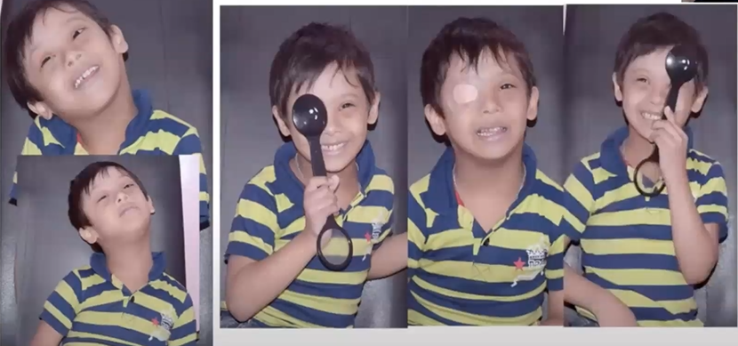
A mother from Indonesia shared her story about her now six-year-old son, who was born with a cleft palate and DS. They regularly took him for eye exams, and when he was four, a doctor found cataracts in both eyes that had not been apparent before. He had cataract surgery in both eyes and had to wear glasses due to myopia. It was a struggle to get him to wear the glasses. In mid-2021, his right eye suddenly became red. They discovered he had total retinal detachment in that eye. Today he is dependent on his left eye.
Another mother shared the difficulty of taking a child with special needs to a physician. This can provoke anxiety in the parent, which she believes rubs off on the children. It is comforting to find a physician to trust who also feels comfortable with your child, especially since this makes the child more likely to want to return.
The last video was of parents with their daughter, who has DS. She said, “I love to sing and dance. I sing and dance very well.”
Dr. Nischal ended the presentation by saying, “Chromosomes don’t care what you look like, what language you speak, what religion you are when you’re affected by DS. It doesn’t take away your aspirations and hopes, and the whole family is affected. One of the things I’m very proud of is that with EEF and the Children’s Foundation in Pittsburgh, we provide a space for these families. Even adult patients with DS come to us at the Vision Institute and at CHP, and we have some amazing professionals that take care of them.”
He then praised a “wonderful coffee shop” in the Strip called Bitty & Beau’s, “a human rights movement disguised as a coffee shop.” Most employees have DS, and it is “an absolute pleasure to buy coffee from them.”
“I am truly privileged to be able to say that I work at Children’s Hospital of Pittsburgh and the VI together with professionals like Dr. Jhanhji to deliver care and hope for patients of all ages with DS,” Dr. Nischal said.