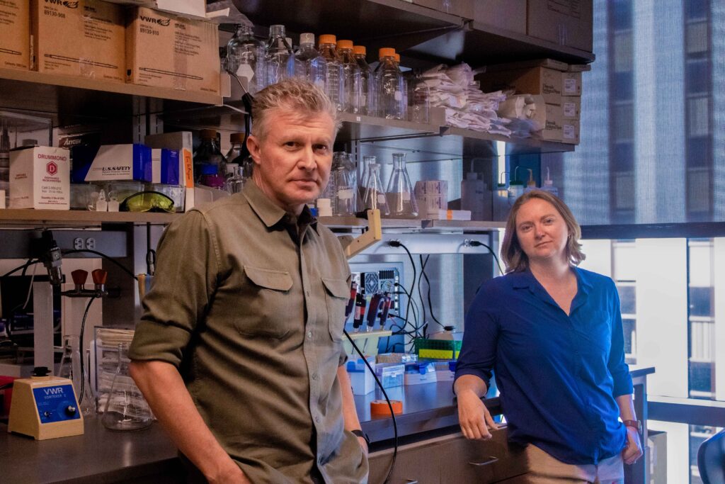
Two new faculty members are collaborating on the structural biology and neurobiology of retinal interconnections. Bryan Jones, PhD, Professor of Ophthalmology, and Rebecca Pfeiffer, PhD, Assistant Professor of Ophthalmology, came from the Moran Eye Center, University of Utah, and started at Pitt on August 1, 2025, continuing the work they started in Utah.

Dr. Jones studied neurophysiology and neurodiagnostics in graduate and post-graduate school and then joined the lab of Dr. Robert Marc where his PhD dissertation work pioneered the understanding of retinal plasticity now known as retinal remodeling. “We pioneered retinal connectomics during my postdoctoral fellowship with Dr. Marc,” he said. “Then working with Dr. Rebecca Pfeiffer in my lab, we pioneered pathoconnectomics.”

Dr. Pfeiffer also worked in Dr. Marc’s lab as a graduate student, where she first became interested in the consequences of retinal degeneration on the inner retina and retinal remodeling. “Retinal remodeling is a well-established phenomenon of plasticity which occurs as a consequence of photoreceptor degeneration, regardless of the precipitating injury,” she explained.
While characterizing the long-term attributes of retinal remodeling, Dr. Pfeiffer became specifically interested in two major attributes: Müller glia metabolic changes that occur early in retinal remodeling and persists throughout the later stages of remodeling and wide-spread neurodegeneration which occurs as the last component of retinal remodeling following long-term photoreceptor degeneration.
“These two primary projects indicated that glia were among the earliest responders to photoreceptor degeneration, even before obvious photoreceptor degeneration had begun, and that the retina is not privileged compared to the rest of the central nervous system and will also demonstrate progressive neurodegeneration when a component of its neural network degenerates,” Dr. Pfeiffer said.
Because of the late-stage neurodegeneration, Dr. Pfeiffer wanted to see how the inner retinal networks were affected across key time points of retinal degeneration. This interest was complimentary to Dr. Jones’ interests and was the basis of her postdoc with him.
“It was previously described, in part by Drs. Jones and Marc, that many neuronal classes of the retina extend aberrant processes during and following photoreceptor degeneration as a primary aspect of retinal remodeling,” Dr. Pfeiffer said. “Investigating neural networks and the precise connectivities that underly them is the main goal of the field of connectomics, in which Dr. Jones is a world expert.”
Together, they decided that the next frontier of connectomics should be pathoconnectomics, or evaluating the network changes occurring in neurological disease. Based on this and the healthy rabbit connectome already available and partially annotated by Dr Marc and Jones’ groups, Drs. Jones and Pfeiffer decided to make a series of pathoconnectomes in a rabbit model of retinitis pigmentosa. These pathoconnectomes range from early retinal degeneration, as rod photoreceptors are degenerating, but not lost, to late in retinal degeneration following the complete loss of photoreceptors and after neurodegeneration is well underway.
“The construction of the three pathoconnectomes and their progressive analysis has demonstrated rod bipolar cells make new contacts with cone photoreceptors prior to the loss of rod photoreceptors and they also make novel electrical contacts in the inner retina at a similar timepoint,” Dr. Pfeiffer explained. “These retained and altered networks (with many more which remaining to be evaluated in these volumes) will help to guide therapies to interface with the degenerate retina and identify obstacles of remodeling needing to be addressed in therapeutic strategies.”
Dr. Pfeiffer said one of the most interesting aspects of connectomics/pathoconnectomics is the sheer quantity of image data that is produced. A single research group could analyze a volume for decades and still not ask every question the data exists to answer. “The technical skill and infrastructure required to generate these volumes is immense and only a few groups worldwide have cultivated the necessary combination, which makes the only logical option to share these rich resources,” she added. “Based on this, we took the somewhat unorthodox plan to continue sharing volumes generated by our groups moving forward to answer fundamental questions about native and diseased circuitry (in the Jones group), and glial connectivity and alterations in disease in mine.”
Dr. Jones said about the work, “We are bringing new software-based approaches to core ultrastructural services which will benefit all investigators in the Department as well as bringing spatial metabolomics approaches. My lab is also extensively collaborative with active projects across the globe.”
He also called Dr. Pfeiffer a superstar. “She is precisely the kind of scientist that will help push science, medicine and our society forward,” he added. “She is curious and will benefit any community she is a part of through her hard work, and dedication to her interests and the people that make up the communities she is a part of. It is also fair to say that her work in my lab was largely responsible for our pioneering work in pathoconnectomics.”
Dr. Pfeiffer said she has never had the opportunity to be part of a Department that truly spans from the basic research, of which she considers herself a part, to translational medicine, working to implement therapies to save and restore vision as soon as is safely possible. She also said the current potential to preserve or restore vision for individuals with retinal diseases has never been higher, so having the opportunity to contribute to this effort is exciting.
Dr. Jones was drawn to the UPMC Vision Institute because of its leadership. “Leadership matters, especially when trying to push the boundaries of science which is what the mission of vision research here is all about,” he said. “Drs. Sahel and Ash have curated a phenomenal group of people here to understand vision and discover therapies for vision loss. This is the right environment where we can push the boundaries of our science to understand how retinas are wired and how retinal circuitry breaks in retinal disease.”