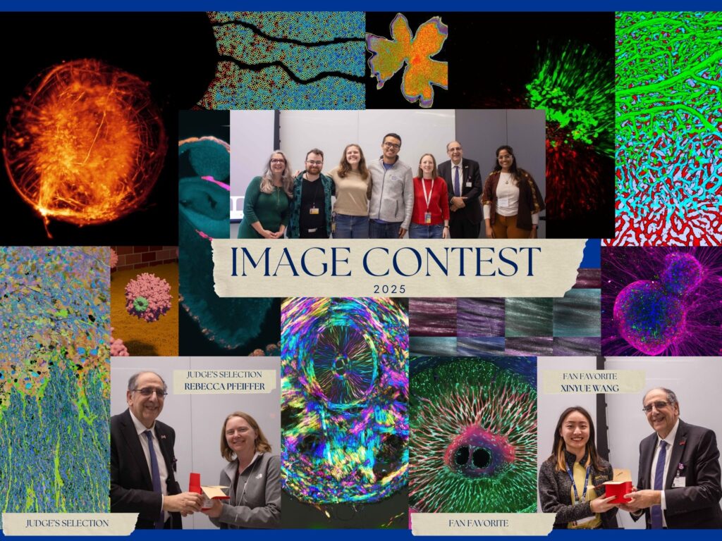
The Mercy Vision Institute Scientific Image Contest recently announced this year’s winners. Out of about 70 submissions, 12 made the shortlist, one clinched the top prize, and another was chosen as the fan favorite.
Leah Byrne, PhD, Assistant Professor, Department of Ophthalmology, started the contest last year to communicate the “amazing breadth of research conducted at Pittsburgh.” This year’s contest was organized by a committee of Department trainees, including Joao Ramos Ferreira, Bhavya Iyer, Sree Motipally, Annu Rani, Michael Ferrone, and Annika Oberdorfer, advised by Dr. Byrne and Tony St. Leger, PhD, Assistant Professor of Ophthalmology and Immunology.
This year’s judges were Nancy Washington, PhD, Eye & Ear Foundation Board member; José-Alain Sahel, MD, Department of Ophthalmology Chair; and Jennifer Lawrence, Director of Exhibitions at the Kamin Science Center.
According to the contest website, the judges were looking for “striking, beautiful, and informative scientific and clinical images related to the field of ophthalmology and vision science.” Images were evaluated on aesthetics, creativity, and demonstration of innovative science.
“The vibrant colors and gentle movement of the images created an intriguing experience,” Washington said. “The contest provided an opportunity for others to experience seeing images from a different perspective. It brings out aesthetic qualities revealed by the tools of scientific research. This makes the experience accessible and meaningful for a broad community.”
Any kind of image was allowed, ranging from microscopy to scientific drawings and even time lapses. The contest is open to anyone working in the field of ophthalmology and vision science at or in collaboration with the University of Pittsburgh.
The shortlist images will be showcased during the year at the UPMC Vision Institute, printed in the Eye & Ear Foundation newsletter, added to the Department’s website, and featured in a monthly calendar and on swag.
Rebecca Pfeiffer, PhD, Assistant Professor of Ophthalmology, was the Top Prize winner. In addition to the above, she received a $100 Amazon gift card and bragging rights. The Fan Favorite image by Xinyue Wang, a Health Sciences Research Fellow, was selected by the audience on the day of the award ceremony and was gifted a $100 Amazon gift card.
Rebecca Pfeiffer
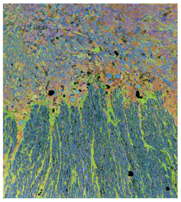
The retina is a multilayered structure consisting of dozens of types of neurons and a few major types of glia. When the light-sensing neurons of the retina (photoreceptors) degenerate, many downstream cells respond through changes in metabolism and neural circuit wiring. In this image, the retina is labeled for the metabolites Taurine (red), Glutamine (green), and Glutamate (blue). Through the combination of these metabolites, we can see the major types of neurons and glia of the retina. The glia take on two distinct appearances. Bright green for the astrocytes running through the blue ganglion cell axons, and yellow, orange, and red for the Müller cells spanning through the rest of the retina. The multicolored Müller cells indicate diverse changes in their metabolism occurring as a consequence of the photoreceptor degeneration. The image is named for the appearance similar to an aerial photograph of waves crashing onto a beach.
Xinyue Wang
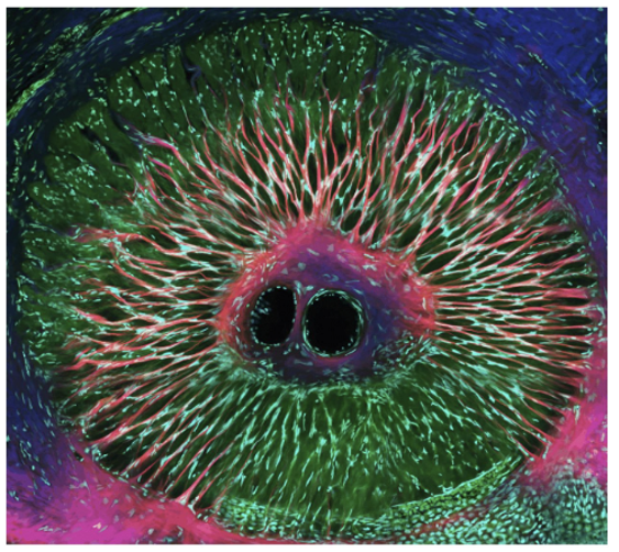
Collagen and cell nuclei in a tree shrew optic nerve head sample.