By John Ash, PhD, Larry Benowitz, PhD, and Emily Woodward
This year, the University of Pittsburgh expanded the focus of its annual Fox Center for Vision Restoration Conference, achieving two goals. The first was to launch the newly opened Gilbert Foundation Validation Research Lab, which is now part of the Fox Center. The program included a half-day of research presentations from local and invited speakers funded by the Gilbert Foundation, focusing on innovative projects related to optic nerve gliomas caused by mutations in the neurofibromatosis 1 gene.
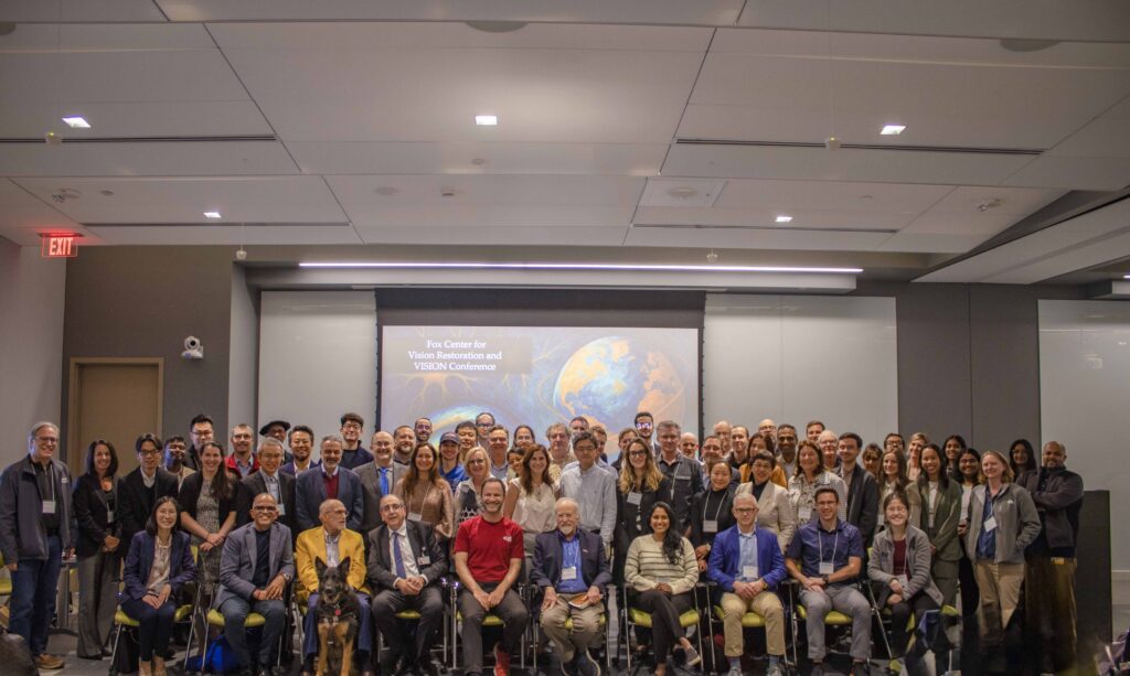
The second was to hold a two-day conference featuring a series of talks from local and invited speakers participating in an ambitious Whole Eye Transplant project funded by the Advanced Research Projects Agency for Health (ARPA-H). Both meetings were combined and took place at the Vision Institute in Pittsburgh from October 27 to 29, with 193 in-person and virtual attendees.
The conferences began with keynote presentations from Petr Baranov, MD, PhD, of Harvard, and longtime colleague of Pitt Ophthalmology Chair Dr. Sahel, Botond Roska, MD, PhD, founding director and senior group leader at the Institute for Molecular and Clinical Ophthalmology Basel and a professor at the University of Basel, Switzerland. Dr. Baranov discussed his research on identifying gene networks that regulate the formation of optic gliomas in Nf1 patients. Dr. Roska outlined a promising new technology involving mitochondrial transplants and other organelles to correct blinding disorders caused by mutations in mitochondrial genes, aiming to regenerate retinal ganglion cell axons. He emphasized the importance of targeting treatments based on specific cell types using molecular binding partners. Additionally, he presented ongoing work on developing treatments that could reverse myopia — the largest vision problem worldwide, affecting over 3 billion people.
The Gilbert Foundation recently awarded research grants to Ophthalmology faculty members Silmara de Lima, PhD, and John Ash, PhD, to support studies on optic nerve gliomas. Both studies are being led by Dr. de Lima, who joined the Department in 2025. The Nf1 research program included 10 presentations from both leading clinicians treating Nf1 patients and scientists working to understand the molecular mechanisms of the disease and developing next-generation therapies.
The whole eye transplant meeting included 38 presentations from investigators from institutions across the United States who are members of the VISION team led by Dr. Sahel and Jeff Goldberg, MD, PhD, of Stanford. VISION scientists presented studies aimed at developing the tools, technologies, and therapies needed to successfully perform a functional whole-eye transplant. This means recovering a donor eye and preserving its function, transplanting it successfully into a new patient, reestablishing the blood supply, and stimulating the neurons that convey visual information from the eye to the brain (retinal ganglion cells, RGCs) to regrow new connections back to the exact the right places in the patient’s brain. The Fox Center for Vision Restoration and VISION Conference featured cutting-edge presentations with pivotal insights on how this goal can become a reality.
VISION scientists also presented their latest breakthroughs in regenerating the optic nerve. The optic nerve is a vital pathway for vision, connecting the back of the eye (retina) to the brain, enabling the brain to interpret signals from the eye into meaningful images. Treatments aimed at protecting retinal ganglion cells and/or regenerating the nerve fibers (axons) of these cells to restore vision included therapies that promote growth factors associated with lens injury and healing, regulating immune cells, utilizing biomechanical signaling, electrical stimulation, nerve wraps, and various forms of gene therapy targeting different cellular mechanisms. The discussion also covered the function of recovered cells, emphasizing not only the need to regenerate the nerve fibers (axons) that transmit visual signals from the eye to the brain but also to ensure they operate effectively to truly restore vision. Scientists also explored potential therapies to ensure the regenerated cells connect precisely to the correct areas in the brain, along with technologies that map the path of the axons to guide them accurately.
Another group of scientists presented their inventions and recommendations for preserving donor eyes after recovery. One major goal of the ARPA-H project is to develop technologies and protocols to successfully keep recovered donor eyes viable for up to 48 hours. Several presenters achieved or nearly achieved an initial 24-hour target, with plans to extend this to 48 hours. Importantly, the presenters emphasized maintaining the functional capabilities of the eye within that period, not just keeping it alive. They measured calcium, oxygen, glucose, lactate, electrophysiological responses, and other indicators to assess how well the eye was surviving. The scientists adapted successful procedures from transplant techniques used with other organs, such as the heart and lungs. They also outlined criteria to evaluate which donated eyes would be the best candidates for transplant, considering factors like cause of death, cell density, and thickness.
In addition to preserving the eye before transplant, scientists at the Fox Center and VISION Conference discussed the most promising protocols for successfully transplanting the eye and preventing it from being attacked by the host’s immune system. Tools used in this effort include nerve wraps and AAV2-based gene therapies. The scientists in this group are using a variety of technologies, including ultrasound, machine learning, electrophysiology, and single-cell RNA sequencing, to map out the immune response after the eye has been transplanted and identify potential targets for therapy. One scientist presented a robotic arm and enhanced imaging system that can provide more detailed feedback to surgeons and execute parts of the surgery in hard-to-reach locations. Additionally, scientists shared approaches that will minimize harm to animals used in studies and preserve the face shape of eye donors.
Underpinning all three components of a successful whole eye transplant – preservation, transplant, and optic nerve regeneration – is the need for comprehensive, clear, and detailed imaging to make it possible for scientists to spot trends and analyze the impact of their proposed treatments. Scientists also shared their success stories and recommended equipment and techniques to use for optical image capture. Adaptive optics was discussed as a groundbreaking tool that is comparable to microscopes in its level of importance to the ophthalmology field. Multimodal imaging and visible light OCT were also discussed, along with the challenges of capturing images from patients during the transplant process.
This year’s meeting was unique in several ways. First, it brought together a large group of investigators with complementary expertise to address two major ophthalmological challenges: Protecting and restoring vision in people with Type I neurofibromatosis, and the enormous task of restoring vision by transplanting the eye of a recently deceased donor into a blind patient. This challenge can only be tackled by assembling teams of investigators with complementary skills in areas such as tissue preservation, microvascular surgery, electrophysiology, gene therapy, immune regulation, functional testing, and the molecular biology of retinal neurons. Such a team has never been assembled before, and the excitement of having these specialists together for two days was palpable. The goals are very ambitious, but if they are achieved, this is the right group to accomplish them.