At the beginning of the Eye & Ear Foundation’s January 21st webinar, “Cranial Base Surgery: The Story So Far…and What’s Next,” Pittsburgh native Garret Choby, MD, said he is thrilled to be back in Pittsburgh. The Associate Professor of Otolaryngology-Head & Neck Surgery with a dual appointment in Neurological Surgery at the University of Pittsburgh School of Medicine was recruited to UPMC in 2023, where he specializes in sinus and skull base surgery. His goal for the webinar was to provide insight into the Cranial Base Surgery Center.
What is Skull Base Surgery?
The question Dr. Choby is asked with some regularity is, “What is skull base surgery?” This can be a challenging one to answer, he said. “To put in perspective, what we’re really focusing on are tumors that arise at the junction of the bottom of the skull and sinonasal cavity,” he added. “We divide these into three different areas: anterior (front) cranial fossa, middle cranial fossa, and posterior cranial fossa.”
When surgeons like Dr. Choby talk about approaching tumors in the nose, they name the surgical approach based on the structures they will be traversing to get there. In the sagittal plane, this includes approaches like transfrontal, transplanum, and transclival. The coronal plane is an approach that extends beyond the middle, outwardly, or laterally along the anterior, middle, and posterior cranial fossa, accessing tumors that arise in these “really critical neuroanatomic areas.”
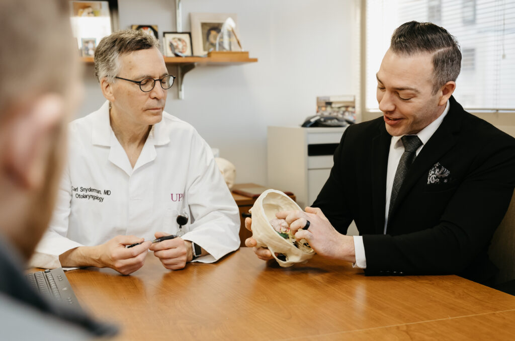
Cranial base surgery is a collaborative team-based surgery that requires a lot of resources and collaboration across departments. The core team at the University of Pittsburgh comprises otolaryngologists and neurosurgeons. Georgios Zenonos, MD, Carl Snyderman, MD, MBA, Garret Choby, MD, and Eric Wang, MD, FACS, form the core of the surgical aspect of skull base surgery. But it extends beyond surgical care, with several critical partners they rely on. In addition to otolaryngology and neurosurgery, they work with oculoplastics, radiation oncology, medical oncology, pathology, and endocrinology.
Historical Shift
Historically, skull base surgery required removal of a large piece of the skull bone, retracting or removing the brain backwards, and accessing the tumor through a narrow corridor to get to the tumor in the deep skull base, otherwise known as an external craniotomy. Now, in a minimally invasive endonasal surgery, surgeons approach select tumors through the nose and arrive at the tumor very early on with excellent visualization.
An external craniotomy means having blind spots, standard instrumentation, and sequential teams. Surgeons go from the outside-in and the tumor is removed last.
Endoscopic surgery involves special instrumentation, the ability to look around corners, and simultaneous teams. Surgeons go from the inside out and avoid the need for brain retraction.
Pittsburgh is a Leader in Skull Base Surgery
Pittsburgh has been a leader in skull base surgery for many years. In fact, Pitt was one of the first centers in the world dedicated to cranial based surgery back in the 1980s. It was the landing ground for the endoscopic endonasal era in the late 1990s and early 2000s.
The number of academic peer-reviewed publications that come from an institution show its impact, Dr. Choby said. Pitt has the most, with about 30% of all publications at the time this study was done.
Pitt is the epicenter for endoscopic skull base surgery (EEA) and has taught others how to do this “Pittsburgh style” surgery through a course held since 2005. There have been over 50 courses.
In 2025, Pitt celebrated its 5,000th EEA, which is “far and away the largest experience in the world.” Pitt does about 250 EEA surgeries a year.
Tumors
Tumors addressed are intracranial tumors and sinonasal tumors. Intracranial means it arises in the brain cavity. Sinonasal tumors are in the sinonasal cavity and spread along the skull base or into the brain area. The most common one the team deals with is pituitary adenoma (intracranial).
A very challenging tumor is clival chordoma, a large cancerous tumor deep in the skull base in the area between the brain stem and sinonasal cavity. Pitt has become one of the leading centers for clival care, receiving a number of referrals annually from around the country and the world.
Meningioma is a relatively common benign tumor that arises from the lining of the brain cavity.
The second large category of tumors addressed via EEA are sinonasal tumors. One such example is olfactory neuroblastoma, which arises high in the sinonasal vault and quickly spreads intracranially. Squamous cell carcinoma is the most common subtype of cancer arising in the sinonasal cavity, frequently in the right maxillary sinus. Sinonasal mucosal melanoma (SNMM) arises in the lining of the sinonasal cavity and is “extraordinarily aggressive.” It spreads quickly and frequently means metastatic.
What makes these tumors challenging?
- Aggressive biologic behavior
- Critical surrounding structures
- Visualization
- Surgical expertise
- Reconstruction of the cranial vault
“As we’ve learned more and have better equipment, we are able to visualize these tumors quite well, often even better than through an external approach,” Dr. Choby said.
If a large tumor is taken out through the nose, the patient is left with a large defect or hole in the skull base, which puts them at high risk for meningitis or infection. With the newer techniques, surgeons have excellent success in reconstructing the skull base and re-separating the brain cavity from the sinus and nasal cavity once the tumor is removed.
In addition to not needing a brain retraction or craniotomy, benefits of an EEA are a shorter hospital stay and improved quality of life. Pitt is one of the main centers studying quality of life outcomes for these tumor types and an EEA approach.
Current and Upcoming Efforts
Dr. Choby outlined the team’s goals:
- Push the envelope of patient care for skull base tumors
- Innovation
- Optimizing outcomes
- Patient-centric
- Collaborative
For sinonasal malignancies, surgeons occasionally “induction” therapy and systemic agents. For squamous cell carcinoma – which often spreads quickly and is in the eye socket—the goal is orbital preservation. The five-year overall survival rate is around 50-55%.
Immunotherapy
PD-1 inhibitor is a newer agent that is part of the immunotherapy cadre. It is a protein expressed among tumor cells that, when bound to the body’s immune system, forces that T cell to die. The T cell is one of the body’s immune system surveillance cells that will attack and destroy cancer cells. Immunotherapy allows the body’s own immune system to survive.
Dr. Choby is surgical co-director for an upcoming clinical trial that is the first NCI trial focused on sinonasal cancers in about 20 years. Pitt is collaborating with UNC, along with other NCI-designated institutions. Enrollment will start in 2026. The trial will targeting aggressive sinonasal cancer with neoadjuvant therapy. People with T3 and T4 – the two highest local stages of a tumor – will be enrolled and randomly either receive neoadjuvant chemotherapy or those two agents with an added PD-1 inhibitor therapy for two cycles, followed by imaging. If there is a good response, the patient will move on to surgery, followed by adjuvant radiation therapy and chemotherapy as dictated by current standard regimens. “I am excited to learn how well these patients can do with neoadjuvant therapy or induction chemotherapy, and when we add in PD-1, what additional benefit that may provide,” Dr. Choby said.
Sinonasal Mucosal Melanoma
This is a unique disease because it is melanoma but in the sinonasal cavity. “This is the ugliest of the ugly tumors,” Dr. Choby said. That means survivability is very poor, which gives them good impetus to study and focus on this disease process. The overall survival rate at five years is around 20-22%. In other words, at five years out, about 80% of patients will have died.
Compared to cutaneous melanoma, sinonasal mucosal melanoma is a more aggressive subtype, with early spread of the tumor to the neck nodes and distantly in the body. From a biologic tumor perspective, it has distinct drivers with a very low tumor mutational burden, which is counterintuitive.
Key non associated factors in whether the patient has a three-year recurrence or metastasis free survival are:
- Perineural invasion
- Tumor depth
- Margin status at surgery
- Tumor stage
- Radiation
- Chemo
Things that do matter are brisk infiltrating lymphocytes, which signal the body’s immune system response to the tumor. Proliferative indices dictate how quickly cells are dividing. The mitotic rate predicts the ability to recur or spread. This is different than many other tumor types. However, the current staging system does not predict this well at all – mitotic or metastasis free survival.
Ki67 is a marker of how rapidly a cell is dividing and appears to be suggestive of survival over time. The presence of tumor infiltrating lymphocytes is a marker of the body’s ability to identify and infiltrate the tumor with its own immune cells.
Immune Checkpoint Inhibition
Immune checkpoint inhibitor therapy is being used more frequently to attack SNMM. In a multi-center international study of SNMM with metastatic disease, looking at ICI vs biochemotherapy (interleukin, interferon), the team found that when ICI is given, overall survival is much better than traditional chemotherapy or immunotherapy.
For neoadjuvant use of immunotherapy, there is minimal data, so three studies (five patients) looking at this published in 2024 showed a response rate of 40%. Early data is pretty convincing in that patients who had a complete or partial response to that neoadjuvant immunotherapy had a much-improved ability to survive later.
Ongoing Initiatives and Future Directions
Pitt is involved in multi-site collaborative efforts, like the CORSICA initiative. Since this is a very rare disease process with only a handful of cases a year, a long-standing effort with nine other centers is studying sinonasal cancer including MM. All patients are prospectively enrolled, prospective oncologic or cancer-related data, including quality of life, is collected. A biobank of all tumor specimens helps improve overall numbers.
The Pittsburgh Skull Base “Think Tank” comprises former Pittsburgh trainees who work together to pool data for rare diseases and answer important questions about clinical care for those patients. Dr. Choby called it a fruitful effort that is paradigm shifting.
Inocras is a start-up company partnered with the CORSICA group, pairing work on a prospective data center and data banking with whole genome sequencing. By sequencing the entirety of the genome, this allows a comprehensive look at all coding and non-coding areas of the genome, which leads to an increased ability to identify mutations. Down the road, this will provide insight into targeted therapy, which is the holy grail.
RNA sequencing is an important area to look at – studying not just genomic data but how it is expressed in regard to RNA and proteins, which will lead to how to better target treatment.
There will be more to come with neoadjuvant use of immunotherapy. Pitt is currently leading a five-site study looking at the utilization of neoadjuvant therapy. More data will likely be published next year.
“For me, what does it come down to?” Dr. Choby asked. “The patient who is sitting in front of me.” He shared two patient cases. One was someone who came from a different state in 2020 with a locally advanced SNMM – a right sided tumor. The main symptoms were vision loss and proptosis (eye pushed forward). There was early spread to lymph nodes in the neck. Five-year estimated survival was less than 4%. Dr. Choby treated him with two agent immunotherapies for two cycles followed by surgery and radiation and one year of single agent immunotherapy. As of 2025, the patient is cancer free with no tumor in imaging and clear lymph nodes. “A truly amazing result,” Dr. Choby said.
On the other hand, a different out-of-state patient came to him in 2022 with a striking similar tumor. The spouse of a physician, she got an appointment as soon as she was diagnosed with a locally advanced right sided tumor involving the right-side orbit and no nodal disease. She was treated the same way as the other patient, but by the time she got to surgery, the disease had metastasized. She died very quickly afterwards.
Why? What is the difference? The first patient was set up for success with low Ki67 and brisk TILs (tumor infiltrating lymphocytes), whereas the second patient had a very high mitotic rate, high Ki67 and weak TILs.
“There is lots of work to do, but we’re excited about the future,” Dr. Choby said.