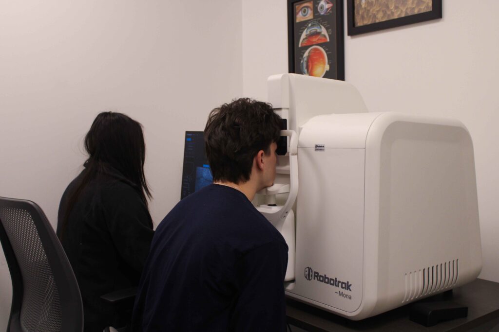
By Emily Woodard, Program Manager, Department of Ophthalmology
At the end of 2025, the Department of Ophthalmology purchased a new prototype human eye imaging system from Ocutek Corporation called the MONA IV, a type of adaptive optics scanning laser ophthalmoscope (AOSLO). This adaptive optics technology provides high resolution imaging in a user-friendly format. It will be maintained by the Rossi lab, but it is available for others in the Department to use after they receive the proper training.
Adaptive optics technology provides more fine-tuned images by using a deformable mirror to correct for ocular aberrations, which are distortions from imperfections in the eye’s optics. The MONA IV machine can capture images at the cellular level, including images of immune cells, bacteria, nerve fibers, axons, capillaries, blood cells, and cone photoreceptors.
Ethan Rossi, PhD, expects to use the equipment to identify potential causes of eye inflammation in patients, which occur in many different ophthalmic conditions. Some work that may be enabled includes identifying infectious pathogens and potentially grouping the imaged immune cells into different classes of cells. Dr. Rossi also expects that this equipment will support his lab’s research on age-related macular degeneration, examining how the condition evolves over time and looking for specific cell signatures to help identify which patients are at the greatest risk for a more severe disease progression.
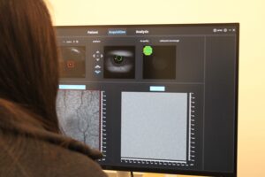
The advantages of this new commercial AOSLO prototype compared with other adaptive optics technologies are numerous. It is easier to maintain than other imaging technology housed in the Rossi lab, and easier for others to learn to use. Additionally, one notable feature of the MONA IV is that it has a built in simultaneous wide field of view for navigation and improved correction over larger refractive errors. This makes it easier to capture images from a wider swath of patients’ retinas and in patients with worse optical quality, including those with conditions like myopia. The wide field of view will allow users to more easily match imaging locations between MONA IV images and clinical images. While the majority of the images taken by the MONA IV will be used for research, they can also help to inform clinical treatment of patients, and the Rossi lab is currently working with several of our ophthalmologists to evaluate the use of this new technology in their patients.
There are currently only two of these machines in the United States, however, Ocutek has customers around the world. Notably, Dr. Rossi’s collaborators at the Institut de la Vision in Paris have also purchased this equipment. Dr. Rossi is very excited about the opportunity that this parallel technology creates for current and future multi-centric studies, including current collaborative scientific investigations and potential future clinical trials.
Later this year, a complementary animal imaging system will also arrive, with the same functionality as the human machine, but adding fluorescence capabilities that will capitalize on the ability of cells to be fluorescently labelled for identification in animal models. Dr. Rossi hopes that this will encourage collaborations where results from animal testing can be compared with the human eye images to better understand and interpret changes seen in patients. Using the same technology for both will allow for “apples to apples” comparisons that can help discern whether results found in animal studies are replicable in humans and whether existing models of disease in animal systems match what is seen in human disease.
Dr. Rossi will also receive an additional upgrade to the MONA IV system with adaptive optics OCT being added later this year for cross-sectional imaging of the retina. These new imaging systems will continue to position the Department of Ophthalmology at the forefront of ophthalmic imaging technologies, facilitating collaborative research projects and enabling cutting-edge discoveries.