The Eye & Ear Foundation’s November 13 webinar, “Choroidal Melanoma: What Do We Know and the Current Treatment Paradigm,” was presented by one of the vitreoretinal surgeons in the Department of Ophthalmology at the University of Pittsburgh. Farzad Jamshidi, MD, PhD, Assistant Professor, talked about one of the more common intraocular tumors – choroidal melanoma.
What is Cancer?
Dr. Jamshidi described cancer as an uncontrolled proliferation of “mutant” host cells that do not respect anatomic boundaries. They grow, invade, and overtake the spaces of normal cells. Almost any cell in the body can become such a “mutant.”
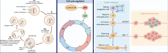
Some of the early critical understanding of cancer biology actually came from tumors of the eye. Years ago, cases of retinoblastoma – a tumor in children that can be inherited — led people to think that perhaps there were genetics behind cancer, leading to understanding one of the key tumor suppressor pathways and cell cycle dysregulation in cancer biology.
Eye Anatomy and Cancers of the Eye
The eyeball, or globe, is important for vision and has different parts and lots of supporting structures. The orbit, or eye socket, also houses supporting tissue to the globe. Cancer can not only happen in the eye, but the supporting tissues can also develop malignancies.
Ophthalmology subspecialities are divided by eye anatomy. As a retina specialist, Dr. Jamshidi specializes in treating diseases of the retina and also deals with intraocular tumors – tumors that happen inside the eye.
Uveal Melanoma
Uveal melanoma is the most common primary intraocular cancer in adults, with about 2000-3000 annual cases in the U.S. It is more common in lighter skin colors. This condition is devastating as there has not been much progress in the past couple of decades in terms of improving survival, but there is research trying to better understand it.
The uvea is comprised of the iris, ciliary body, and choroid in the middle layer of the eye. The outside of the eye is the sclera. The retina is like wallpaper inside the eye that captures images and sends to the brain via the optic nerve. The middle vascular pigmented layer that supplies the retina is the choroid. Melanomas can occur in any of the uveal segments, but the most common is the choroid.
Precursor Lesion
A choroidal nevus is like a mole on the skin, but in the eye. It is benign and a lot of people have it. While these are not cancer, in about 1 in 8,000 cases, the cells accumulate mutations, which start dividing and disrespecting the boundaries of the eye, structure, and body, hence turning into choroidal melanoma.
Choroidal Melanoma
Choroidal melanoma has a variable five-year mortality rate depending on its genetics and presentation. With regards to its size, if it is small, the mortality rate is 1-6%. If medium, the rate is 15-20%, and if large, it is 35 to >50%.
Of these cases, 85% spreads to the liver, which is a vital organ, and poor in bearing metastatic burden and is really what leads to mortality. About 20-30% of people experience spread to the lungs.
Other than metastases, complications can include growth inside the eye overtaking structures of the normal eye leading to a blind, painful eye with elevated pressure. This extraocular extension is a bad sign when seen in patients.
Goals of Treatment
The goals of treatment are survival and life, keeping the eye, and retaining as much visual functionality as possible. “Generally speaking, we want to treat and recognize tumors early,” Dr. Jamshidi said. While these are slow growing tumors over a period of months, once there is evidence of growth, that means it is abnormal and should be treated.
Unfortunately, many treatments eventually do affect vision depending on the tumor’s location. Sometimes people do extremely well if the vital structures of vision are not adjacent to the tumor.
Treatment: Enucleation
Historically, the main treatment for large tumors was to remove the eye. A technique that has been around for a long time is enucleation or removing the eyeball and a section of the optic nerve. It is replaced with an implant to create space, and then a prosthetic is placed on the eye.
Ocularists in Pittsburgh do an amazing job with the prosthetic; they paint the prothesis to match the other eye. It can be hard to tell whether the eye has been removed.
Treatment: Eye Salvage
An ophthalmologist in the UK named H.B. Stallard did pioneering work in the 1930s. He had patients that lost an eye who were losing the other eye to the same cancer. He came up with an idea to sew radioactive seeds to the outside of the eye, where the radiation would kill the tumors.
The process has advanced since then. Now there is a different version of the same concept, called plaque brachytherapy. It involves little gold plates – almost the size of a bottlecap – filled with radioactive seeds. Different seeds are used; the most common one in the U.S. is iodine 125. The plate is placed where the tumor is, so the radiation kills the tumor. The plate has a gold cover to block the radiation going backwards.
In order to place the plate, it is sutured on the back of the eye. Radiation is therefore localized, and damage elsewhere is minimized.
Surgeries are two parts. In the first one, the plaque is placed onto the eye. Collaboration with colleagues (radiation physicists and radiation oncologists) helps calculate the dosage. When the tumor is dead – usually after a couple of days – the plaque is taken out.
Brachytherapy Surgery
Because the eye media is made of clear tissue, surgeons can look inside and see the tumor and mark it. First a clear plaque is placed to mark the suture locations. Sometimes other structures around the eye have to be moved to create space, like removing muscles and then returning them in the second surgery.
Not all tumors can be treated with this technique. It is particularly useful for medium sized and smaller tumors. Larger ones unfortunately mean removing the eyes or using external radiation. If the tumor surrounds the optic nerve 360 degrees, plaque therapy cannot be done.
External Radiation Treatment
Another kind of treatment is externally delivering radiation to the eye, not via a plaque. This can be applied to larger tumors as well. In proton beam therapy, a proton beam is used to kill the tumor; it looks almost like a column of radiation.
Another technique, with expertise here in Pittsburgh, is gamma knife surgery, which involves collaborating with neurosurgery colleagues. The day of the procedure, the eye is numbed by the ophthalmology team, preventing it from moving around during the procedure. A “halo” is fixed to the skull. “All this fixation is very important to make sure we deliver radiation exactly where we want it,” Dr. Jamshidi said. This is done in cases where plaque cannot be used.
Challenges in Treatment
Even in quickly recognized choroidal melanoma cases, early micrometastasis occurs. Does the immune system play a role in holding these at bay before they become life-threatening growths? Despite its rarity, however, uveal melanoma can shed light onto cancer metastasis in general.
Novel Therapies
Currently there is ongoing research, with great people working trying to improve patient outcomes. An interesting trial that is now analyzing its data looked at viral particles that preferentially bind to choroidal melanoma and only get activated with a certain wavelength of light. Dr. Jamshidi called this a “very interesting” concept to treat this cancer earlier.
Future Work
At the UPMC Vision Institute, Dr. Jamshidi with Dr. Ethan Rossi and colleagues are doing vascularity studies with advanced imaging to help distinguish nevi from melanoma.
“In short,” Dr. Jamshidi concluded, “choroidal melanoma is the most common primary intraocular tumor in adults. We do have treatment options, and they work better than no treatment, but there is a lot of room for improvement still.”