Scientists in the Department of Ophthalmology at the University of Pittsburgh School of Medicine continue to advance their research on a variety of areas that could eventually help solve the puzzle of vision loss. One current area of focus involves studying the fovea.
What’s a Fovea?
The fovea is a small depression in the retina of the eye where visual acuity is highest. The center of the field of vision is focused in this region, where retinal cones are mainly concentrated. The fovea is responsible for our ability to see colored and sharp vision and perform daily tasks, like reading, driving, and recognizing faces. Thus, any detriment to this area has a significant impact on our sense of sight and our quality of life. Although highly relevant to our high acuity vision, our understanding of the molecular mechanisms regulating fovea formation is just starting to be revealed, given that most traditional laboratory model systems lack a fovea in their retinas. Indeed, foveas are only present in some primates among mammals, in a few species of fish and lizards, and interestingly in a large number of birds.
Egg-ceptional Findings
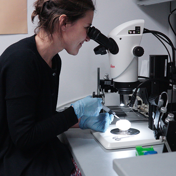
Led by Susana da Silva, PhD, Assistant Professor of Ophthalmology, researchers are studying the development of chick embryos to better understand the development process of the human eye. “We have shown that the chick retina possesses a central high acuity area (HAA) comprising many of the same foveal hallmark cellular features as humans, such as complete absence of rods, high density of specialized slender cone photoreceptors, increased number of cells in the inner nuclear layer and the highest density of ganglion cells,” states Dr. da Silva. Her team has identified the first molecular mechanism that mediates HAA development in chicks, and importantly, has found similar patterns of development in human embryonic development, suggesting a common mechanism between humans and birds.
Why Did the Chicken Cross the Road? … For Progress
This breakthrough validates chickens as a model for studying fovea development, paving the way for potential direct implications for humans. Chick embryos offer a nearly perfect study specimen, as they are very accessible, amenable for embryological techniques, and can be acquired at high numbers. Dr. da Silva’s team is currently performing a number of experiments, some of which are aimed at understanding if and when the development of the fovea in humans and chickens differ. “The overarching goal of my research is to advance our understanding of the basic genetic and molecular mechanisms underlying fovea development and subsequently establish new experimental models of human retinal diseases affecting the fovea with wide applicability and therapeutic potential,” says Dr. da Silva.
What Dr. da Silva is learning will provide clinicians and scientists with an understanding of how care can be advanced for conditions such as Age-Related Macular Degeneration, Glaucoma, Optic Nerve, and Retinal Regeneration. This type of innovative research is made possible with support from generous donors to the Eye & Ear Foundation. If you would like to be a part of this groundbreaking work being done, please consider making a gift to help support our research.
For the latest news on ophthalmology research, subscribe to EEF’s Monthly Newsletter.
