Bryan W. Jones, PhD, retinal neuroscientist and Professor of Ophthalmology at the University of Pittsburgh brought his lab over from the University of Utah. In the Eye & Ear Foundation’s September 24th webinar, “Retinal Circuitry and Retinal Degeneration,” Dr. Jones said the move has been wonderful. He has traveled to ophthalmology departments around the world and the one at Pitt that Dr. José-Alain Sahel, MD, and Dr. John Ash, PhD, have built is like no other. Having 63 research faculty is “phenomenal,” he said, but more important than the number of research faculty is the quality, “not only as scientists but as human beings.” Dr. Jones described the faculty as incredibly accomplished, wonderfully collaborative, and collegial.
He then went on to share the research from his lab. First, he showed an image of the sun, captured every day by the Nasa Solar Dynamics Observatory. This image illustrates that our eyes – what we use to see – have evolved because of the photons or light that comes off the star.
Next, Dr. Jones described eye anatomy. The front of the eye houses the cornea, the clear structure that is operated on during surgeries like LASIK to improve vision. The iris is next and is like an aperture; it gives eyes their color. Behind that is the lens, which gets operated on or replaced when we have cataracts. For most of us who live long enough, this will be replaced at some point. The retina is at the back of the eye, a combined layered, cake-like structure with sensors at the back and layers underneath. If you look in the eye, the retina is transparent and almost looks like wet tissue paper when it is operated on or dissected.
The Retina
The vision research community has been studying retinas for over 130 years. Ramoni Kahal in 1892 understood that the retina processed information and that retinal neurons are directional and computational. However, while he understood that these neurons were wired together, modern neuroscientists did not understand for a long time just how complex these connections are. There have been a lot more recent modern anatomical techniques that are now used to understand these tissues and figure out how they are wired together.
The retina is a wonderfully complex tissue with many cellular superclasses classes and many more subclasses of glial, horizontal, vascular, amacrine, bipolar, and ganglion cells. Each one of these neurons has a unique relationship with the retinal network associated with it. “We’re interested in figuring out how information flows from the photoreceptors,” Dr. Jones said. When a photon flows through the retina and hits the photoreceptors at the back, how that information is propagated through the retina and its circuitry is like a highly parallel supercomputer that gives us a sense of vision.
Most of us have a rod pathway of vision that functions at low light levels – the rod axon and rod photoreceptor. This is the visual pathway that allows us to see at night or in starlight. When the moon starts to come out and we can start to see color in the world, that is when these photoreceptors — the red, green, and blue ones — or cones start to turn on. When it comes to the cone photoreceptors, these are the photoreceptors that let us see color in the world. Most of us can see red, green, and blue, but about 8% of us are color blind; we only see two or three colors. Because the gene for the opsin, the molecule that actually detects the photons, is carried on the X chromosome, those people with two X chromosomes have a tiny percent chance of being tetrachromatic or have the ability to see four colors rather than three that most of us see.
It turns out that there are also visual and non-visual components of vision that we are only recently beginning to understand, with a substantial portion of our retinal ganglion cells that are themselves photosensitive and do things like set our circadian clock.
“The cool thing about retinas is they give us a number of opportunities to study how these neural systems are wired, how they function, and how they undergo degeneration because of simply how accessible they are in experimental conditions, particularly with animal models,” Dr. Jones said.
Studying the Wiring of Retinas
So, how does one study the wiring of retinas? Standard microscopes cannot be used; a special one called an electron microscope is used. The connections between neurons are smaller than the wavelength of light, so to see these connections, you cannot use microscopes that use photons.
People have been using electron microscopy for some time. Helga Kolb – “one of the gods of vision science” – was one of the first people to use this to study the retina intensively and use 3D reconstruction to study the connections between neurons. She also founded the world’s first textbook called Webvision, which is used by people around the world to train students and physicians on the retina. Dr. Jones took over managing it in 2000 as webmaster and editor and then last year when he moved to Pitt, he brought it with him.
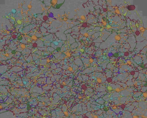
Creating a complete wiring network map has been a challenge in the field. Figuring out how to represent the image data and have it make sense from a circuit diagram perspective was another challenge. In just one example, one photoreceptor might have 17 separate connections. Multiply that by 50, and that results in 51 separate cells. “You need some way to describe the complexity of the network, and the problem is it turns out that even describing these simple networks over time can get a little complex,” Dr. Jones explained.
The next step was to look at some of the math and literature behind networks. Dr. Jones shared the thought journey and explained how the process of mapping these circuits with ultrastructure became known as a field called connectomics. This is the precise study of how these neural networks interact and how they are connected with one another, which offers road maps to network topologies.
As Dr. Jones explained, each slice in the “layer cake” that makes up the retina was made at 70 nanometers thick. That is literally thinner than the wavelength of light. If the section is turned sideways, it disappears. But this section can be put under an electron microscope, where thousands of separate images are made into a mosaic.
A number of databases have been built this way. The first was a rabbit retinal connectome. A mouse retinal connectome has been built, as has a non-human primate. A human retinal connectome is being worked on now. Each of these data sets is around 16-90 terabytes in size and takes well over a year, sometimes two years, just to capture and build. While complex, this is teaching how neural systems work and maybe how to build biologically inspired silicon-based computers, which is important because biological systems are way more efficient than silicon-based systems in how they process information.
“The take-home message is that retinal circuitry is way more complex than we thought,” Dr. Jones said. “And another surprising take-home message from our analyses is that retinal circuitry is also way more precise than we thought.”
Dr. Jones acknowledged the software architect who has made this all possible, because special software is needed to view and analyze this data.
How this Work Impacts Retinal Degenerative Diseases
“Here’s where the rubber meets the road for disease with this kind of work,” Dr. Jones said. A whole bunch of disease processes rob people of their vision. His PhD dissertation was centered around how the retina changes in retinal degenerative disease, and it turns out that the circuitry of the retina is fundamentally altered in disease. Understanding how retinal degenerative diseases from retinitis pigmentosa to AMD, glaucoma, optic nerve atrophy, and other forms of blinding disease or trauma that impact millions globally is driving this work.
Dr. Jones said they wanted to know how the wiring in retinas was changing in disease by examining the synaptic connections.
A few years ago, Rebecca Pfeiffer, PhD, did a post-doctoral fellowship in Dr. Jones’ lab. She built three pathoconnectomes – the first in the world using a transgenic rabbit they engineered that has a form of autosomal dominant retinitis pigmentosa. Analyses of this data is still ongoing. The first set they are analyzing is very early in the stage of retinal degeneration while there are still photoreceptors present. One thing they discovered is that there are some incredibly precise, stereotypical, and predictable changes that start happening to the wiring of degenerating retinas.
The neat thing about the software is the ability to take cells from the databases, render them out in three dimensions, move them around, and then click on the connections between the neurons. The location of that connection can be inputted into the ultrastructural database. Researchers can then go to exactly the spot where the neurons connect. By doing this, instead of expecting a chemical synapse in one example, they found a different kind that functions very differently – a gap junction.
Effectively, what is happening is two circuits that are involved with low light vision are completely rewiring, turning on our ability to detect when light levels go down. This throws the system into conflict. These gap junctional changes are seen in AMD and retinitis pigmentosa. There is good evidence that it also happens in glaucoma.
One of the first things that happens in many retinal diseases is when patients tell their doctors that it is taking them a long time to adapt from low light environments to bright ones or vice versa. “We think these changes in gap junctions with rod bipolar cells and off bipolar cells and A2 amacrine cells is the molecular/cellular reason behind this complaint,” Dr. Jones said.
The other discovery is that retinal circuitry fails in more predictable ways than previously thought. “This is pretty cool and it’s pretty exciting because it tells us that there are targetable interventions that we may be able to target using gene therapy or potentially pharmacologic therapies to slow down this process of degeneration,” Dr. Jones said.
Many of the gap junction changes and protein changes being explored in the retina happen in Alzheimer’s disease, opening up the possibility of using retina models of degeneration as a tool to explore other kinds of neural degeneration earlier in the course of disease than previously possible. One of the problems in the Alzheimer’s community is that everyone waits until the very late-stage disease when a lot of damage has already occurred, complicating the ability to treat.
Another current project has to do with the 13-line ground squirrel. When this animal hibernates, the photoreceptors in their retina degenerate and the synapses come apart looking just like they do in human retinal degeneration. But when they come out of hibernation, they reform those synapses and regrow their photoreceptors. During their hibernation, they are resistant to glaucoma as well. There are lots of questions about this, so stay tuned.